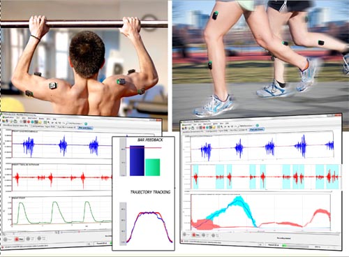
(a) sEMG Surface electromyography (sEMG), also known as dynamic electromyography (DEMG), is a bioelectrical signal when the neuromuscular system is guided and recorded from the muscle surface through electrodes. It has a different degree of correlation with the active state and functional state of the muscle, and thus can reflect the activity of the neuromuscular to some extent. The bioelectricity generated in the muscle movement generates a potential difference through two measuring electrodes (relative to the reference electrode), and after the differential amplifier detects the signal, the image obtained after amplification and recording, the modern high-grade sEMG is an amplified signal. It is then converted into a digital signal and transmitted to the microcomputer through the communication system. The analysis software in the microcomputer analyzes and processes the obtained data to complete scientific research or clinical diagnosis tasks such as test evaluation. sEMG is a simple, non-invasive, electrophysitic activity that is easily accepted by subjects. It can be used to test a wide range of EMG signals and to help reflect changes in muscle physiology, biochemistry, etc. during exercise; The quiescent state measures muscle activity, and can continuously observe changes in muscle activity during various movements; it is not only a diagnostic evaluation method that is meaningful for motor function, but also a better biofeedback treatment technique [1]. Therefore, in the diagnosis of neuromuscular diseases in clinical medicine, the ergonomic analysis of muscle work in the field of ergonomics of colleges and universities, the fatigue determination of sports systems (physical institutes), the rationality analysis of exercise techniques, the types of muscle fibers and the absence of anaerobic thresholds. Injury prediction, neuromuscular disease diagnosis in hospital rehabilitation, and evaluation of muscle function have important practical value [2]. (two) sEMG commonly used analysis indicators Frequency domain analysis The analysis method is to perform fast Fourier transform (FFT) on the sEMG signal, obtain the spectrum or power spectrum of the sEMG signal, reflect the variation of the sEMG signal in different frequency components, and better reflect the change of sEMG in the frequency dimension [3]. At present, the following two indicators are commonly used in frequency domain analysis, namely, mean power frequency (MPF) and median frequency (MF). The MF slope has been used as a fatigue index during the maintenance of isometric shrinkage. The following physiological phenomena can occur during muscle fatigue: such as synchrony of motor units, changes in the order of recruitment of slow/fast muscle fibers, changes in metabolism (including changes in the form of energy production, hypoxia, increased H + concentration, cell membrane conductivity) Lower), the frequency of the EMG signal tends to shift at a lower frequency. Therefore, the EMG signal can be used for fatigue measurement and the physiological phenomena associated with the fatigue process can be measured. SEMG can obtain indicators on fatigue (or endurance testing) through a fast spectrum analysis program [4]. In addition, the FFT spectrum curve of sEMG is not a typical normal distribution. Therefore, from the statistical point of view, using MF to describe the spectral characteristics of sEMG is better than MPF, but in practice, it is found that the activity reflecting muscles MPF is more sensitive in state and functional state. 2. Time domain analysis Time domain analysis is to treat the EMG signal as a function of time. It is used to characterize the amplitude characteristics of time series signals, including integral electromyography (IEMG), root mean square (RMS), and mean amplitude (MA). Integral myoelectricity refers to the sum of the area under the curve per unit time after the obtained myoelectric signal is rectified and filtered, which can reflect the change of the myoelectric signal with time [5]. EMG is used to analyze the contraction characteristics of muscles in unit time [6], and its calculation formula is as follows: iEMG=∫t+Tt|EMG(t)| dt The root mean square value (RMS), like IEMG, can also reflect the variation of the amplitude of the sEMG signal in the time dimension. It is directly related to the electrical power of the EMG signal and has a more direct physical meaning. The mean amplitude (MA) reflects the strength of the muscle electrical signal, and the number of participating motor units. 3. Joint analysis of EMG spectrum and application (JASA) JASA analysis [7] was first proposed by Alivin. Luttmann et al., a new fatigue measurement method that considers EMG amplitude and spectrum changes simultaneously, so as to better reflect the real situation of muscle fatigue. The amplitude-frequency combined analysis simultaneously considers the amplitude and spectral changes of the sEMG signal to effectively distinguish similar phenomena of changes in myoelectric signals due to increased muscle strength or fatigue. 4. Wavelet analysis The wavelet analysis method [8] is an analysis method that combines the time domain and the frequency domain. It has a variable time domain and frequency domain analysis window, which provides a reliable way for real-time processing of signals. Through the appropriate wavelet transform for the EMG signals under different functional states, the frequency change and time characteristics can be observed at different scales. Some studies have suggested that wavelet analysis is suitable for the analysis of unsteady EMG signals in athletes. By using the time-frequency positioning characteristics of the wavelet transform, the time-varying spectrum analysis of the signal can be realized, and the signal can be analyzed in any detail without being sensitive to noise. Therefore, wavelet transform is a powerful tool for surface EMG signal analysis. (III) Research status of sEMG in muscle function assessment 1. Clinical evaluation of neuromuscular lesions using sEMG Electromyography is an important part of neurophysiological testing. Muscle testing can be used to distinguish between neurogenic and myogenic damage and the extent of damage and to detect new potentials and functions, thus providing an accurate and detailed objective basis for clinical use. The clinical scope for muscle diagnosis is as follows: (1) Myogenic diseases (muscle fibers): including various types of chronic progressive muscular dystrophy, polymyositis, myotonic syndrome, congenital myotonia, atrophic myotonia. The relationship between myoelectric integral value and muscle strength and muscle tension is: there is a positive correlation between the myoelectric integral value measured by the surface electrode and the muscle strength when the muscle contracts with static force; the myoelectric integral value is positively correlated with the muscle tension [9] ]. Therefore, the surface electromyogram can be used to evaluate the changes of the injured neuromuscular function and the difference between the healthy side and the contralateral side, and can be used to observe the progress of the neuromuscular function of the affected side before and after treatment and to formulate and adjust the next treatment plan accordingly. [10]. This program has application value in the evaluation of muscle strength, muscle tone and muscle fatigue in dysfunction caused by neuromuscular diseases in rehabilitation medicine; sEMG plays an important role in the diagnosis and differential diagnosis of myotonic myopathy, and can be diagnosed by sEMG. Congenital muscle rigidity, atrophic muscle rigidity, congenital accessory muscle rigidity. (2) Neuromuscular junction disease: myasthenia gravis, myasthenia gravis syndrome. EMG repeated electrical stimulation attenuation wave phenomenon can not only help to confirm the diagnosis, but also help to identify the classification of myasthenia gravis. The latter's low-frequency stimulation amplitude is attenuated by more than 10%, but the high-frequency stimulation is greatly increased. However, some scholars believe that EMG can only make classified diagnosis for myasthenia gravis, and must be combined with muscle biopsy and initial clinical diagnosis, otherwise it may lead to misdiagnosis of such diseases. (3) Qualitative and localized diagnosis of diseases such as muscular atrophy, numbness, weakness, and physical activity disorder for unknown reasons. After muscle atrophy, the cross-sectional area of ​​muscle fibers decreases and the muscles contract continuously due to unloading. The muscle power spectrum changes after muscle atrophy. sEMG provides a new method for non-invasive detection of muscle atrophy. For example, the abnormal nerve conduction velocity of electromyography is one of the diagnostic criteria for amyotrophic lateral sclerosis (ALS). The occurrence rate of CMAP amplitude/DML×F wave is an effective objective electrophysiological index, which can be used for ALS conditions and The prognosis was quantitatively assessed. (4) Evaluation of the efficacy of rehabilitation therapy. For muscle rehabilitation treatment, especially rehabilitation training methods, it can be used as a method for evaluating the comparison of pre- and post-treatment effects and follow-up. For example, sEMG is used to diagnose the paraspinal muscle function in the diagnosis of low back pain. In the case of surgery, trauma, neck and shoulder pain, and other muscle dysfunction, determine the severity of muscle dysfunction, pain, etc. through potential changes in myoelectric signals. SEMG can be combined with other advanced rehabilitation tests and training equipment to assist Diagnose and evaluate various conditions affecting skeletal muscle function, such as combining with video images to better analyze the actions of certain daily activities; and combining with gait analysis systems to analyze the myoelectric activity of abnormal gait; The synchronous camera system combined with the control study helps to analyze and correct various abnormal gaits; it is more clear with the balance test trainer and the constant velocity test system. (5) Myopathy of other diseases, etc. The modern SEMG test system can extract the EMG signal and use the display and the horn to feedback the image signal and the sound signal to the subject respectively, realize the dual-signal feedback therapy, enhance the training effect, and greatly help to improve the muscle strength. It can be used for the treatment of various muscle atrophy and spasms. Special electrodes can also be used to detect and train pelvic floor muscles for the prevention and treatment of urinary incontinence, uterine prolapse and hemorrhoids. 2. Application of sEMG in measuring muscle fatigue The measurement of muscle fatigue is of great significance in both rehabilitation medicine and sports research. In 1975, Petrofsky et al. proposed that the center frequency (CF) of the myoelectric power spectrum shifts from high frequency to low frequency when muscle fatigue occurs. When fatigue causes work to stop, the center frequency is After reaching the same final value, the central frequency has been widely used for quantitative analysis of muscle fatigue. Studies have shown that CF shifts to low frequencies during muscle fatigue and has a good correlation with muscle fatigue. The corresponding center frequency drop curve when the maximum contraction force (MCV) drops by 50% is more sensitive to fatigue and more reflective of fatigue. In terms of the mechanism of muscle fatigue, current studies have confirmed that the ratio of the maximum H wave to the maximum M wave amplitude of eEMG is significantly reduced after the voluntary contraction of muscle contraction. This phenomenon indicates the excitation of spinal motor neurons during muscle fatigue. Sexuality is inhibited, and decreased excitability of spinal motor neurons may be an important factor in the development of muscle fatigue. Through the comparative analysis of sEMG and eEMG, the abnormal effects of muscle fiber activity potential, neuromuscular junction conduction insufficiency, and low motor coil excitability are important causes of muscle fatigue. "Electromyographic fatigue threshold" (EMGFT), established by Matsumoto et al., believes that as muscle fatigue occurs and develops, iEMG and RMS linearities increase, and muscle performance is evaluated. According to a study of 21 female college students, Matsu-moto et al. found that the subjects completed 150W, 200W, 250W and 300W intensity respectively, and the integral EMG of the extra-femoral muscle was compared with the 20-seat treadmill exercise. The exercise time is linearly correlated. The slope of the iEMG curve is linearly related to the load intensity at all levels of motion. It is considered that the application of sEMG can accurately detect the fatigue threshold of the body motion. Some scholars believe that predicting the fatigue threshold of muscles is dynamic or static. In general, with the occurrence and development of exercise muscle fatigue, the FFT curve of surface EMG signals can be shifted to different degrees to the left, and the spectrum curve is reflected. The characteristic MPF and MF produce corresponding declines, and thereby use sEMG to judge the functional status of the muscle. Regarding the reason for the left shift of the sEMG power spectrum, some scholars have explored the relationship between the sEMG power spectrum change and H+ during muscle fatigue, and found that the MPF showed a linear decrease in the biceps muscle during the 60% MVC static fatigue load. After the fatigue load During the recovery period, the MPF recovery was extremely rapid. After only 2 s after the end of the exercise, the MPF had recovered to 26.5% of the entire range of decline; to 30 s, the MPF had recovered to 87.7% of the entire range of decline. From this, it was concluded that: The decrease in the action potential conduction velocity of muscle fibers caused by the increase in H+] is not the only factor determining the left shift of the sEMG power spectrum, suggesting that the left shift of the sEMG power spectrum may be related to the central mechanism of neurogenicity. 3. Use sEMG to evaluate the coordination of muscle strength and muscle activity sEMG provides an important basis and evaluation method for the study of sports science. It can measure muscle strength indirectly during exercise, and can also analyze sports techniques. The application mechanism is that the greater the muscle contraction intensity, the greater the amplitude of the electromyogram. Some scholars use the surface biphasic induction method to detect the EMG and apply the video trajectory system to perform three-dimensional analysis of the three squatting movements. Investigate the strength training of three different squat postures in the normal squat posture, the wide-foot distance squat posture, and the hip joint extension squat posture, and record the effects of the electromyogram reaction and the ground reaction force of the main motor muscles of the lower limbs. Analyze from a physiological and mechanical perspective. It is concluded that the lower limb strength training using the hip joint squatting action plays a certain role in improving the sprinter's ability to improve the running. The method of using the super-equal contraction in the initial motion method is used to improve the muscle explosiveness of the sprinter. effective. 4. Use sEMG to predict muscle fiber type The sEMG signal is the sum of many muscle fiber activities in a certain range. The basic theoretical basis for predicting skeletal muscle fiber type by sEMG is that there is a linear negative correlation between the characteristics of some surface EMG signals (mainly MPF) and the type I fiber ratio during the resistive load. Or positively correlated with the ratio of type II fibers. Most scholars have concluded that those with high fast muscle fiber composition have higher MPF, and the fatigue is significantly decreased, while those with higher slow muscle fiber composition are not significantly reduced [25]. In short, sEMG provides an important basis and evaluation method for the research of clinical rehabilitation medicine and sports science because of its advantages of non-invasiveness, locality and real-time. With the rapid development of computer application and technology, myoelectric diagnosis Technology will also be constantly updated and its scope of application will be more extensive. (reproduced)
Laser Distance Meter module is mainly designed for laser distance
measurer. As the soul part of a laser rangefinder, our module can assure quick
respond, high accuracy and long range measuring.
With
different range measuring program, our modules can satisfy customers` different
requirements, 40m, 60m, 80m, 100m, 120m, 150m, every distance can do multiple
functions, like: Height/Distance/Area/Volume/Pythagorean Measurement. Voice, Bluetooth,
angle measuring, beep, and any other functions can be customized.
We
have been in this line for 10 years, with a strong R&D ability and hard
working, we are now a leading supplier of laser distance meter modules in
China.
Laser Distance Sensor,Laser Range Module,Laser Distance Meter Module,Laser Rangefinder Module Chengdu JRT Meter Technology Co., Ltd , https://www.rangingsensor.com
Analyze how surface EMG is applied to muscle function assessment
