CT measurement of white fat, brown fat and fatty liver in mice API Powder (Active Pharmaceutical Ingredient) Active Pharmaceutical Ingredients refer to the raw materials used in the production of various preparations. They are the effective ingredients in the preparations. They are various powders, crystals, extracts, etc., prepared by chemical synthesis, plant extraction or biotechnology, but Substances that the patient cannot take directly.
According to its source,Active Pharmaceutical Ingredients are divided into two categories: synthetic chemical drugs and natural chemical drugs.Chemical synthetic drugs can be divided into inorganic synthetic drugs and organic synthetic drugs. Inorganic synthetic drugs are inorganic compounds (extremely elements), such as aluminum hydroxide and magnesium trisilicate used to treat gastric and duodenal ulcers; organic synthetic drugs are mainly composed of basic organic chemical raw materials, through a series of organic Drugs made by chemical reactions (such as aspirin, chloramphenicol, caffeine, etc.).
Natural chemical drugs can also be divided into biochemical drugs and phytochemical drugs according to their sources.Antibiotics are generally made by microbial fermentation, which belongs to the category of biochemistry.In recent years, many semisynthetic antibiotics are the combination of biosynthesis and chemosynthesis.Among APIs, organic synthetic drugs account for the largest proportion in variety, yield and output value, which is the main pillar of chemical pharmaceutical industry.The quality of API determines the quality of preparation, so its quality standards are very strict. All countries in the world have formulated strict national pharmacopoeia standards and quality control methods for its widely used API
Active Pharmaceutical Ingredient,API powder, minoxidil PYSON Co. ,Ltd. , https://www.pysonbio.com

Identification of brown fat and white fat
From PLoS ONE, May 2012
Background: Modern lifestyles lead to high levels of obesity. Obesity caused by imbalances in energy intake and consumption is often accompanied by symptoms such as high blood pressure, dyslipidemia, and coronary heart disease, which ultimately leads to abnormal metabolism. The distribution of fat, not the total amount of fat, determines the state of metabolism. Subcutaneous fat is beneficial to the human body, and increased visceral fat and abnormal fat mass in the liver, skeletal muscle and pancreas increase the risk of type 2 diabetes. Another closely related to metabolic diseases is the widespread non-alcoholic fatty liver disease. Recently, brown fat (BAT) has gradually attracted widespread attention. The study of brown fat is mainly concentrated in small animals. However, new data indicate that BAT also plays a role in adults. The amount of BAT is negatively correlated with BMI, indicating the role of brown fat in energy metabolism in humans.
The current gold standard for detecting abdominal fat and liver fat content is MRI and CT. The body fat of mice is usually measured by quantitative magnetic resonance (QMR), which is accurate in measurement, does not require anesthesia of animals and is fast, but does not distinguish between subcutaneous fat and visceral fat, so in this experiment We used LaTheta LCT-200 (Hitachi-Aloka, Tokyo, Japan) to distinguish between abdominal fat and subcutaneous fat and to quantify the two parts of fat. We also used the instrument to measure liver fat content and brown fat.
METHODS: We used different models of lean and obese mice (C57BL/6, B6.V-Lepob, NZO) to determine the optimal scanning parameters for scanning fat at different sites. The data is compared to the actual weighing after scanning. The amount of liver fat is determined by biochemical analysis.
RESULTS: The correlation between the weight of the adipose tissue and the CT measurements was: subcutaneous fat (r2 = 0.995), visceral fat (r2 = 0.990), total white fat (r2 = 0.992). In addition, the use of the abdomen Scanning of the area (between lumbar vertebrae L4 and L5) and body fat can reduce scan time and reduce radiation and anesthesia to experimental animals.
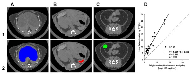
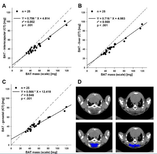
The amount of liver fat obtained by CT scan was linearly correlated with the results of biochemical analysis (r2 = 0.915). In addition, CT measurements of brown fat were highly correlated with the results of the balance measurements (r2 = 0.952). Short-term freezing (4 ° C, 4 hours) resulted in a change in brown fat content, which caused less triglyceride, which was reflected by an increase in CT values ​​during CT imaging.
Conclusion: LCT200 is reliable and accurate for 3D imaging and quantitative analysis of total fat, subcutaneous fat, visceral fat, brown fat and liver fat in mice. This non-invasive method allows us to conduct long-term scanning studies on obesity in mice.
Figure 1. Quantification of hepatic fat by CT. Selected areas of liver (A; blue), spleen (B; red) and WAT (C; green) for determination of mean HU
Upper panel (1): raw gray scale scan slices, lower panel (2): selected organ parts used in calculation of liver fat. (D) Relationship between
The amount of intrahepatic fat isolated and quantified with biochemical analysis and estimations by computed tomography. Dashed line – identity line,
R2 - coefficient of determination.
Figure 2. Brown adipose tissue. Correlation between resected brown adipose tissue (BAT) weighted on scale and estimations of fat depot
Weights by CT. (A) BAT depot in situ (interscapular), (B) resected BAT depot inserted under the liver, (C) resected BAT depot inserted in gonadal fat
Deshed; dashed line – identity line, r2 - coefficient of determination. (D) Analysis examples of two different slices of interscapular brown adipose tissue
Depot by ImageJ (NIH) program. Upper panel: raw gray scale scan slices, lower panel: manually outlined and selected BAT in ImageJ (NIH).
