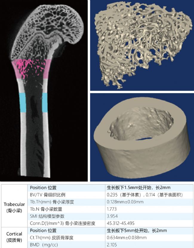
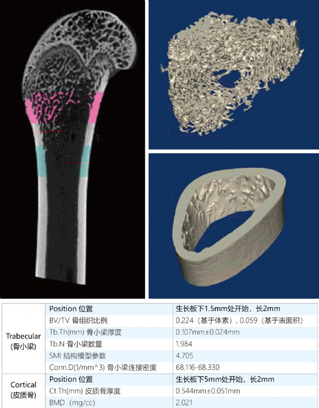
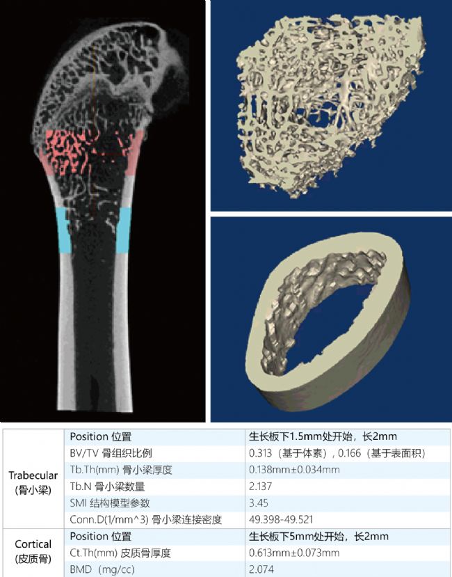
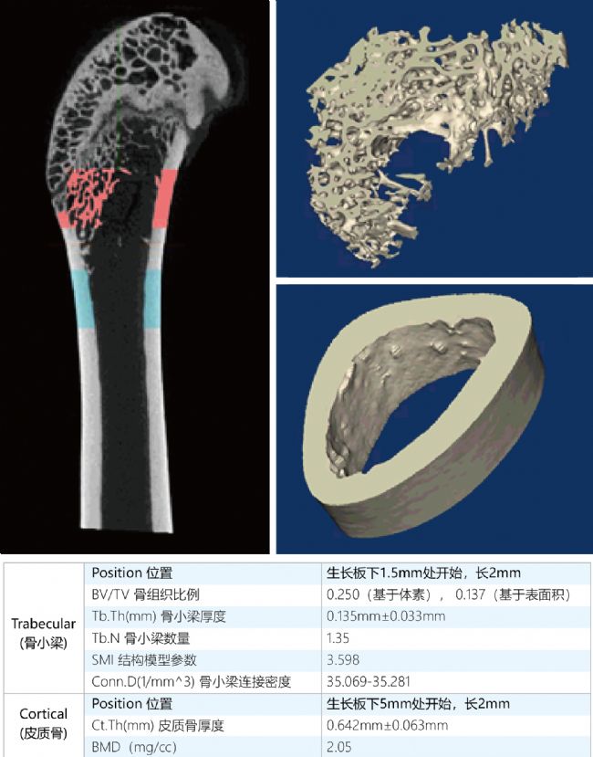
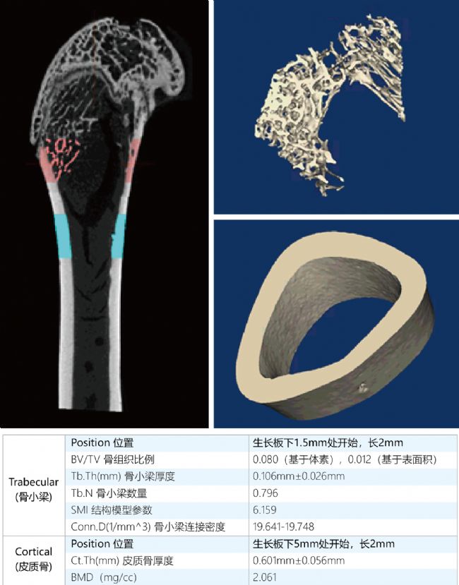
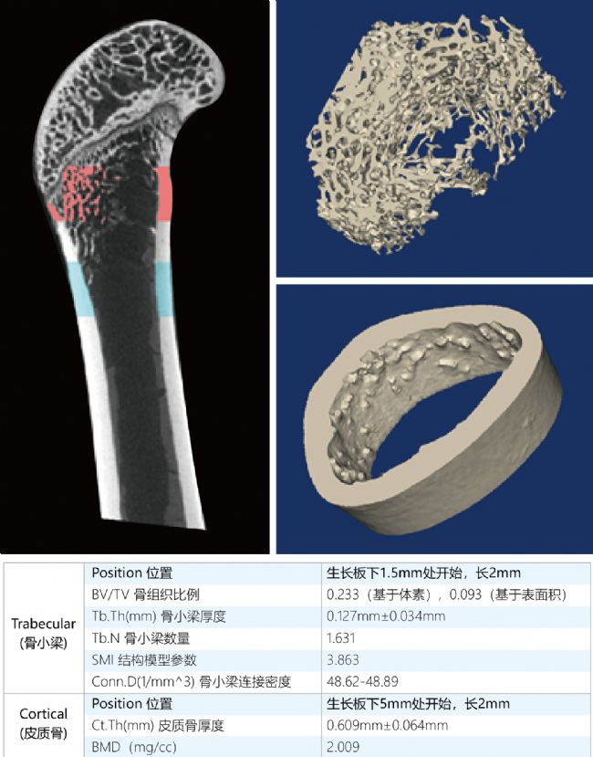
First, the experimental background Estrogen plays an important role in regulating bone metabolism and maintaining the stability of the internal environment. Low estrogen is considered to be the main cause of osteoporosis. The ovaries are organs of egg development and hormone production that are dually regulated by the hypothalamic and pituitary gonadotropins {follicle estrogen (FSH) and luteal auxin (LH)}. Bilateral ovarian ablation in female rats is a more mature animal model that can successfully establish an animal model of osteoporosis induced by estrogen deficiency. Based on this model, it also provides an experimental basis for the development and evaluation of drugs for the treatment of osteoporosis. Second, the purpose of the experiment Osteoporosis is a metabolic bone lesion characterized by a decrease in the amount of bone tissue per unit volume. In this experiment, osteoporosis was induced in rats by ovarian removal, and different drug interventions were given. Micro-CT was used to observe and analyze the degree of osteoporosis after drug treatment (ie, the change of bone tissue volume) to evaluate different drugs for osteoporosis. Efficacy. Third, the experimental process Scanned samples: normal rat isolated femur (group A), osteoporosis isolated femur (B-F group) A is the control group and B-F is the experimental group (the results under different drug treatments) Treatment method: bone trabeculae starting at 1.5 mm below the growth plate and 2 mm in the growth plate of each scanning sample (rat femur), and cortical bone starting at 5 mm below the growth plate and 2 mm long were selected for bone parameter analysis. Modeling method: female rat ovariectomized Acquisition parameters: 60kV, 100μA, FDK reconstruction method Imaging software: Avatar 1.3 (Healthy Medical) Experimental equipment: NEMO ® Micro-CT (Medical Health) NEMO ® Micro-CT (Ultra-high resolution, combined with in-vivo experiments) Fourth, the experimental results Control group (group A) Results of different drug treatments in group B-F Experimental group B Experimental group C Experimental group D Experimental group E Experimental group F V. Experimental conclusion In this experiment, the effects of different drugs on the amount of bone tissue can be seen by the images reconstructed by Micro CT scan, and the relevant parameters of trabecular bone and cortical bone were quantitatively analyzed. The establishment of this experimental model can effectively evaluate the efficacy of different drugs on osteoporosis. Remarks: In the bone analysis related to the large mouse experiment, since the trabecular bone tissue of the mouse bone is a microstructure, the high resolution of the Micro CT device used is the key to the experiment. Acknowledgement: Shaanxi University of Traditional Chinese Medicine conducted experiments and data sharing on the Micro CT equipment of Pingsheng Company. For more case sharing, please pay attention to the public account Life, born for scientific research Website: E-mail: Automatic Massage Foot Bath Machine
The automatic massage foot bath machine is a device that uses water and massage rollers to provide a relaxing foot massage. It usually has a basin filled with water and has built-in massage rollers that move and massage the feet. The machine may also have other functions, such as heat therapy, air bubbles, and vibration. Users place their feet in the basin and the machine provides a soothing massage that helps relieve tension and improve foot circulation. Some automatic foot massage machines also come with removable attachments for additional massage options.
Automatic Massage Foot Bath Machine,Foot Spa Massageer,Heated Foot Spa,Foot Bath Massage Basin Huaian Mimir Electric Appliance Co., LTD , https://www.mmfootspa.com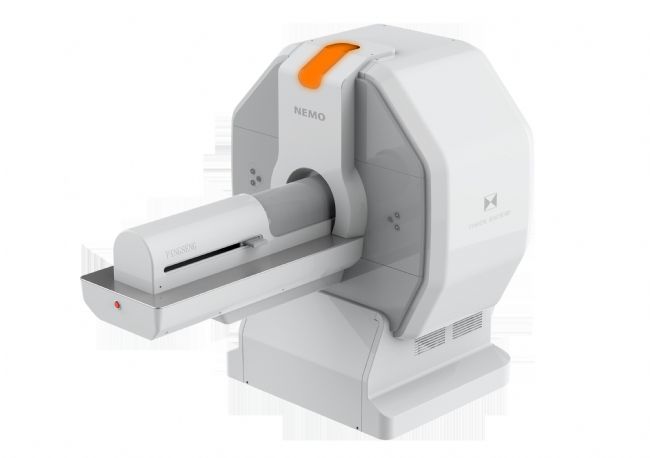

Using Micro CT to explore the efficacy of different drugs on osteoporosis
