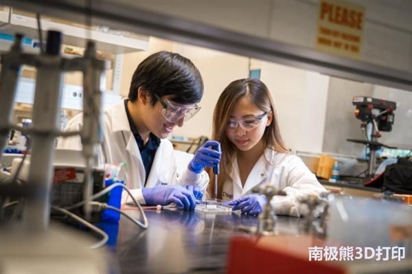
On December 4, it was learned from foreign media that bioengineers at the University of California, San Diego developed an easy-to-use 3D bioprinting technology that uses realistic materials to create realistic organ tissue models. As a proof of concept, the University of California, San Diego's 3D printed vascular network maintains breast cancer tumors in vitro and is a model of vascularized human intestinal tract. (L-R): Bioengineering student Michael Hu and undergraduate Xin Yi (Linda) Lei use their team's new 3D bioprinting technology to build a vascularized bowel model The researchers said that the goal of this study is not to create artificial organs that can be implanted in the body, but to create models of human organs that are easy to grow and can be studied in vitro or used for drug screening. Michael Hu, a Ph.D. student in bioengineering, the first author of the study, said: "We want to make everyday scientists easier - they may not have the specialization needed for other 3D printing technologies - make 3D models of any human tissue they are studying, models It will be more advanced than standard 2D or 3D cell culture and more relevant to humans when testing new drugs, currently on animal models." To create a living vascular network, the researchers first used Autodesk to digitally design a stent. The researchers used a commercial 3D printer to print the stent with a water-soluble material called polyvinyl alcohol. They then poured a thick coating of natural material onto the stent, allowed it to solidify and solidify, and then flushed the stent material into the interior to form a hollow vascular access. Next, they cover the inside of the channel with endothelial cells, which are cells that line the inside of the blood vessels. The final step is to pass the cell culture medium through the blood vessels to keep the cells alive and growing. Blood vessels are made from natural substances found in the body, such as fibrinogen (a compound found in blood clots) and Matrigel, a commercially available form of the actual mammalian extracellular matrix. However, finding the right material is one of the biggest challenges, said co-author of the study, bioengineering undergraduate Xin Yi (Linda) Lei. “We want to use natural materials instead of synthetic materials, so we can make as close as possible to the materials inside the body. They also need to be able to use our 3D printing method.†Ami Dailamy is a graduate student in bioengineering at the Mali laboratory who designed a 3D printing stand In one set of experiments, the researchers used printed blood vessels to keep breast cancer tissue in vitro. "Our hope is that we can use our system to create tumor models that can be used to test anticancer drugs in vitro," Michael Hu said. They extract tumor fragments from the mouse and then embed some of the debris into the printed vascular network. The other fractions were kept in standard 3D cell culture. After three weeks, the tumor tissue encapsulated in the vascular imprint remained alive. At the same time, most of those in standard 3D cell culture have died. In another set of experiments, the researchers created a vascularized bowel model. The structure consists of two channels. One is a rectal tube lined with intestinal epithelial cells to mimic the intestine. The other is a vascular channel (lined with cells) that hover around the intestines. The goal is to reconstruct a bowel surrounded by a network of blood vessels. Each channel is then provided with a medium optimized for its cells. In two weeks, it began to show a more realistic form. For example, the intestines have begun to germinate fluff, which is a tiny finger-like projection on the inside of the intestinal wall. "Through this strategy, we can begin to build complex, long-lived systems in an isolated environment. In the future, this may replace animals used to make these systems, which is what we are doing now," Mali said. “This is a proof of concept that shows that we can organize different types of cells together. It is important if we want to simulate multiple organ interactions in the body. In one print, we can create two different local environments. Each environment maintains different living cell types and puts them close enough so that they can interact," Hu said. This work was recently published on Advanced Healthcare Materials. Future work will focus on optimizing printed blood vessels and developing models of vascularized tumors that are closer to mimicking the body. Sample Release Reagent Kit Of COVID-19 The Sample Release Reagent Kit is intended for the pre-treatment of the samples to be tested. The substances to be tested in the specimens can be released from other substances to facilitate the use of in vitro diagnostic reagents or instruments. DNA release reagent,RNA release reagent,viral release reagent,virus release reagent,sample release reagent kit Shenzhen Uni-medica Technology Co.,Ltd , https://www.unimedicadevice.com

Protein structure is rapidly destroyed by denaturation and biochemical reagents, releasing the nucleic acid.
University of California Screening Drugs with Natural Materials 3D Bioprinted Tissues
