Modular lighting system with ultimate flexibility and expandability The Nikon Ti-LAPP system provides modular illumination for total internal reflection (TIRF), photoactivation/conversion, light stimulation and epi-fluorescence. For individualized research, each module has the flexibility to build a dedicated microscope system. For example, multiple TIRF modules can be integrated into one microscope for different experiments and high speed, multi-angle TIRF imaging. Combined with Nikon TI's layered optical structure, a microscope can be combined with up to five illumination modules (eg, two sets of TIRF, FRAP, DMD, and epi-fluorescence devices). Ti-LAPP modular lighting system | DMD Module Multi-point simultaneous photoactivation The new DMD module enables light activation or light conversion anywhere in any shape. Traditional FRAP components can only be photoactivated with a single point of manual adjustment. Customize the shape, size, position and number of DMD lighting using NIS-Elements software. Researchers can use this technique to label a portion of a cell population or a portion of a protein in a single cell or track the migration of multiple cells. The DMD module is an excellent choice for optogenetic experiments, and optogenetic techniques can alter some cellular or protein functions with light. The DMD module can use a laser as a light source or a smaller phototoxic LED illumination device. Mouse embryonic fibroblasts co-transformed mCherry-labeled Lamin A (red) with the lower left light-transformed light-sensitive GFP-labeled Lamin A (green), which was performed by a DMD module using a 405 nm LED light source. Time series images are performed using fluorescent illumination. By photoactivation of a region of Lamin protein, you can observe the dynamic behavior of local regions. Image courtesy of Drs. Takeshi Shimi and Bob Goldman, Northwestern University Medical School | H-TIRF Module Realize automatic TIRF adjustment and observation The H-TIRF module is equipped with a gradient natural light filter that moves into the light path to make the TIRF illumination uniform. Image courtesy of Melissa Hendershott and Dr. Ron Vale, University of California, San Francisco The bilayer membrane structure prepared in vitro was labeled with Alexa 488 (green) and Alexa 561 (red) and two-color imaging (two-color TIRF) using an H-TIRF illuminator. The proteins aggregate into clusters and exhibit a dot-like structure (upper row). The lower row shows 488 channels and is displayed in a rainbow format to clearly distinguish the difference between the images. The H-TIRF illuminator is used for automatic adjustment of incident focus at different wavelengths. Image courtesy of Drs. Xiaolei Su and Ron Vale, University of California, San Francisco | FRAP Module Analysis of intracellular protein dynamics Mouse embryonic fibroblasts express mCherry-lamin A, and the FRAP module was used in the upper right corner region for spot photobleaching and to study the dynamic changes of lamin A molecules. Time series images use epi-fluorescent illumination. Image courtesy of Drs. Takeshi Shimi and Bob Goldman, Northwestern University Medical School | TIRF Module For cell membrane dynamics and single molecule observation Tetanus Toxoid Vaccine,Toxoid Vaccine,Hep B Immune Globulin,Immunoglobulin Injections FOSHAN PHARMA CO., LTD. , https://www.pharmainjection.com
.png)
 The angle of incidence of the laser observed by TIRF is different from that of the focus as the sample and conditions are observed. Adjusting the angle of incidence and focus to achieve TIRF illumination requires some skill and experience. The new H-TIRF module automatically adjusts focus and angle of incidence by detecting the reflected light speed. These auto-tuning actions are done through the auto-collimation feature in the NIS-Elements software. The penetration angle of the incident angle and the evanescent area can be separately saved and arbitrarily called in different experiments to satisfy the consistency of shooting.
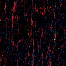
  Fluorescently labeled microscopic (TRITC and Alexa 647) and microscopically bound proteins prepared in vitro were imaged in three colors using an H-TIRF illuminator with a graded ND filter. The angle of incidence of different wavelengths is automatically adjusted.
  If you do not use a gradient ND filter, the TIRF illumination will show an intermediate bright and dark imagination in the field of view. Uniform illumination is achieved with a graded ND filter.
  Using the FRAP module, experiments such as photobleaching, photoactivation, and light conversion can be combined with high frame rate, high sensitivity sensor detection. This module provides point-stimulation of specific regions of the target cell, providing a cost-effective solution for dynamic study of intracellular proteins without a point-scan confocal microscope.
Japan Nikon NIKON inverted microscope multi-module laser application system introduction
Inverted microscope multi-module laser application system Ti-LAPP modular lighting system
